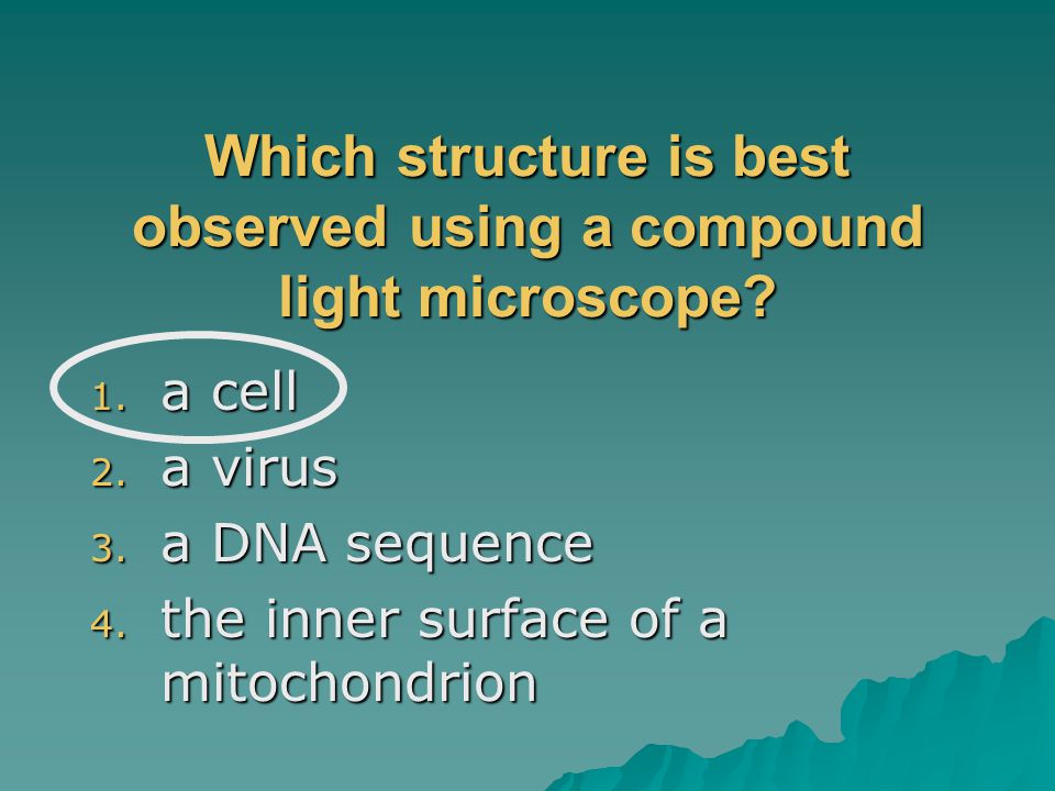
Which Structure Is Best Observed Using a Compound Light Microscope
Compound Light Microscope Optics Magnification and Uses With Links to MicroscopeMaster Buyers Guides A compound light microscope is a microscope with more than one lens and its own light source. 1tissues 2organ systems 3organelles 4organs.
Microscope Definition Parts Functions Types Diagram Uses
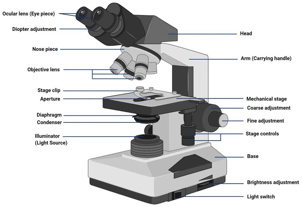
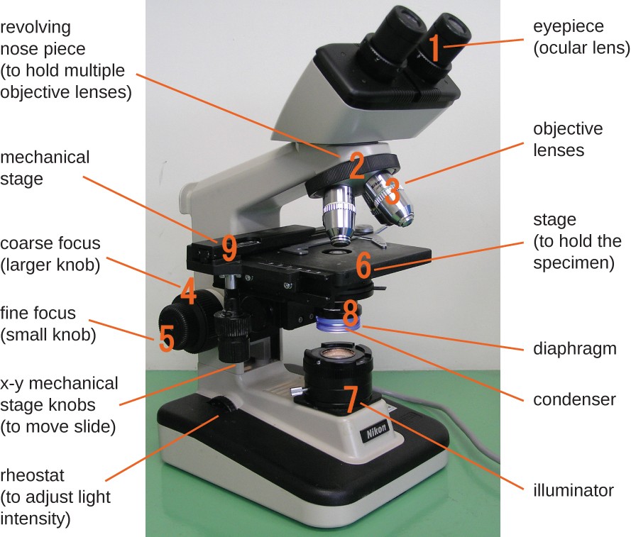
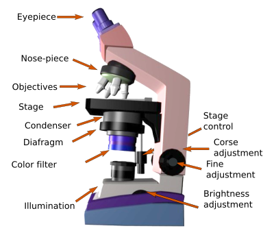
Eyepiece lens or Ocular.
. A student observed several cell layers positioned on top of one another in the high-power field of view of a compound light microscope. The stage micrometre is used to calibrate the eyepiece scale which is then used for measurements. The compound microscope is mainly used for studying the structural details of cell tissue or sections of organs.
A stereo microscope is a type of optical microscope that allows the user to see a three-dimensional view of a specimen. A microscope is considered compound when it has two sent of lensesthe ocular lenses and objective lenses. The base is also known as the foot which is either U or horseshoe-shaped.
The viewer spins the nosepiece to select different objective lenses. Light microscopes magnify the image of the specimen using light and lenses. Iii It is relatively small in size.
The SPO website is best viewed in Microsoft Explorer Google Chrome or Apple Safari. On the rim of the eyepiece there are certain markings such as 5X 10X 15X etc. Cell structure that directly controls regulates the movement of molecules into and out of the cell.
The illuminator is the light source for a microscope. 1 a cell 2 a virus 3 a DNA sequence 4 the inner surface of a mitochondrion. A compound light microscope has its own light source in its base.
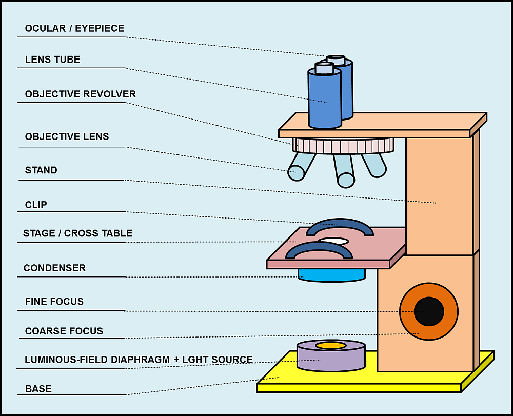
The parts of a compound microscope can be classified into two. A micrometre can be used to properly determine the size of objects seen using a compound microscope. The term compound means that this microscope passes light through the specimen and then through two different lenses.
Using a light microscope one can view cell walls vacuoles cytoplasm chloroplasts nucleus and cell membrane. B Optical Parts of Compound Microscope. Which structure is best observed using a compound light microscope.
Parts of Compound Microscope. The ocular is the lens nearest the eye of the observer. The objects magnified image can be observed with the help of an eyepiece.
Which indicates the magnification power. Which structure is best observed using a compound light microscope. When you use a compound light.
At the top of the body tube a lens is planted which is known as the eyepiece. Easy to use and simple to handle. - Animal cells have many small vacuoles while plant cells have big vacuole.
Nosepiece is a rotating turret that holds the objective lenses. Light microscopes use lenses and light to magnify cell parts. In this type of microscope there are ocular lenses in the binocular eyepieces and objective lenses in a rotating nosepiece closer to the specimen.
A compound light microscope mostly uses a low voltage bulb as an illuminator. The objectives are the lenses nearest the stage of the scope. It is worth remembering that while a good quality microscope will last a lifetime it is a sensitive scientific instrument that will suffer.
The Compound Light Microscope. A compound light microscopes use lenses and light to magnify cell parts. Moving and Placement.
The advantages of using compound microscope over a simple microscope are. O the inner surface of a mitochondrion O a cell O a DNA sequence O a virus THIS IS. I High magnification is achieved since it uses two lenses instead of one.
6Which structure is best observed using a compound light microscope. However they usually can achieve a maximum of 2000x magnification which is not sufficient to see many other tiny organelles. The structure of a cell nucleus would be seen in the greatest detail by use of an electron microscope when using a compound light microscope the most common reason for staining a specimen being observed is to.
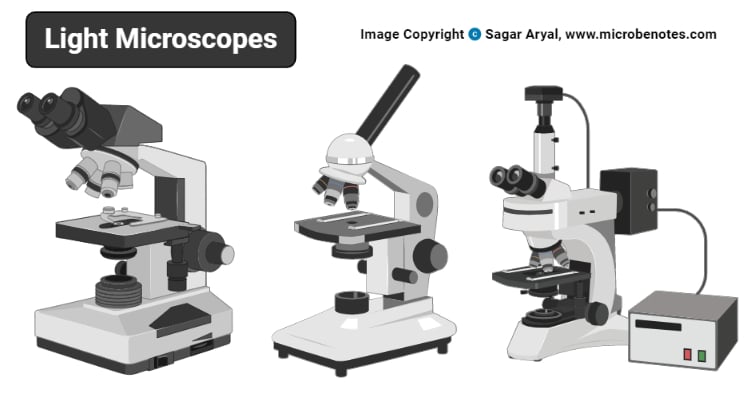
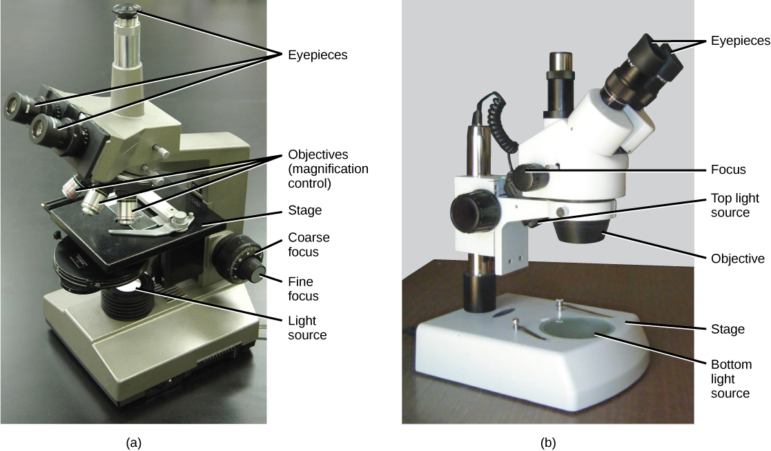
Otherwise known as a dissecting microscope or stereo zoom microscope the stereo microscope differs from the compound light microscope by having separate objective lenses and eyepieces. How to Use a Compound Microscope Familiarization First familiarize yourself with all the parts of a microscope so that you can easily move from one part to another during operation. The stage is the flat platform where the slide is placed.
- Plant cells have cell walls and chloroplasts. However they usually can achieve a maximum of 2000x magnification which is not sufficient to see many other tiny organelles like ribosomes endoplasmic reticulum lysosomes centrioles Golgi bodies unless they have an electron microscope with increased magnification. The eyepiece scale also known as graticule or ocular and the stage micrometre scale make up the latter.
The incandescent light from the light source is reflected by a condenser lens beneath the specimen and the light passes through the specimen up to the objective lens then the projector lens sends the magnified image onto the eyepiece. The image from a light microscope is presented in color. The lens closest to the specimen is called the objective lens while the lens nearest to the users eye is called the ocular lens or eyepiece.
It can be observed with the eye directly recorded by photographic video or computer techniques and image components can be analyzedUsing an objective of NA 14 and green light of wavelength 500 nm the resolution limit is 02 μm. 1organs organism cells tissues 2organism cells organs tissues 3cells tissues organs organism 4organism organs tissues cells 7Which sequence shows a decreasing level of complexity. Differences between plant and animal cells.
- Animal cells have centrioles for cell division. Ii It comes with its own light source.

What Is A Compound Microscope New York Microscope Company
Instruments Of Microscopy Microbiology

Compound Microscope Parts Labeled Diagram And Their Functions Rs Science
1 5 Microscopy Biology Libretexts

Light Microscope Definition Principle Types Parts Labeled Diagram Magnification
Light Microscope Definition Principle Types Parts Labeled Diagram Magnification

Compound Microscope Parts Labeled Diagram And Their Functions Rs Science
Aim How Have Microscopes Ppt Video Online Download
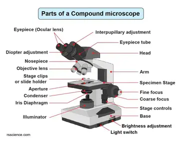
Compound Microscope Parts Labeled Diagram And Their Functions Rs Science
Parts And Components Of Light Microscopes Light Microscope
3 1 How Cells Are Studied Concepts Of Biology 1st Canadian Edition

Light Microscopy Biology Encyclopedia Cells Plant Body Process Animal Different Hormone Used Structure
Histological Techniques 6 Visualization Light Microscope Atlas Of Plant And Animal Histology

Compound Microscope Parts Of Compound Microscope

Light Microscopes An Overview Sciencedirect Topics

Practices Of Science Microscope Use Manoa Hawaii Edu Exploringourfluidearth

Microscope Definition Parts Functions Types Diagram Uses

Optical Microscope An Overview Sciencedirect Topics

Lesson Explainer Microscopy Nagwa


Comments
Post a Comment